Diffusion MRI fiber tracking uses local fiber directions measured at each voxel to track the trajectory of a white matter pathway If we set region A as the seed region and region B as the ending region, does this find tracks ending in both regions?Extract Time Series from Seed Region ¶ For each subject, you want to extract the average time series from the region defined by the PCC mask To calculate this value for sub001, type fslmeants i sub001 o sub001_PCCtxt m/seed/PCC_bin The withingroup functional connectivity analysis for controls revealed a nearsignificant positive correlation in brain activity between the right auditory seed region and the right IFG/pars orbitalis and significant positive correlations between the left auditory seed region and the left IFG/pars orbitalis, as well as the right auditory cortex at the PT/HG border In amusics, brain

Brain Structure And Function In School Aged Children With Sluggish Cognitive Tempo Symptoms Journal Of The American Academy Of Child Adolescent Psychiatry
Seed region mirna
Seed region mirna- Seedbased functional connectivity, also called ROIbased functional connectivity, finds regions correlated with the activity in a seed region In seedbased analysis, the crosscorrelation is computed between the timeseries of the seed and the rest of the brain (telling us where the traffic is communicating between selected cities) (Fig 3, the results are visualized with Tumor and Edema region present in Magnetic Resonance (MR) brain image can be segmented using Optimization and Clustering merged with seedbased region growing algorithm The proposed algorithm shows effectiveness in tumor detection in T1 w, T2 – w, Fluid Attenuated Inversion Recovery and Multiplanar Reconstruction type MR brain images


Decreased Hand Motor Resting State Functional Connectivity In Patients With Glioma Analysis Of Factors Including Neurovascular Uncoupling Radiology
Select initial seed points in MRI images, the hippocampus structure has similar gray values with its adjacent anatomical structures and it is only a small region In order to reduce the computation and improve the segment reliability, we only process a local area, including the whole hippocampus and little other region The first parameter was cerebral and cerebellar asymmetry measured with magnetic resonance (MR) volumetry The second parameter was the functional connectivity from the right and left amygdalae, analyzed on the basis of PET measurements of regional cerebral blood flow (rCBF) during rest and passive smelling of unscented airThe seed point can be manually selected by an operator or automatically initialised with a seed finding algorithm Then, region growing examines all neighboring pixels/voxels and if their intensities are similar enough (satisfying a predefined uniformity or homogeneity criterion), they are added to the growing region "Unsupervised MRI
In this paper, a segmentation system with a modified automatic Seeded Region Growing (SRG) based on Particle Swarm Optimization (PSO) image clustering will be presented The paper is focused on Breast MRI Tumour Segmentation Using Modified Automatic Seeded Region Growing Based on Particle Swarm Optimization Image Clustering Springer for Research & DevelopmentSegmentations of relevant anatomical structures such as breast region and fibroglandular tissue are required Most of state of the art methods for breast segmentation on MRI are semi or fully automated, furthermore they can be groupedincontourbased,regionbasedandatlasbasedapproaches9GenerSegmentation of brain MRI in an image sequence is one of the most challenging problems in image processing, while at the same time one that finds numerous applications In this paper, we propose a robust multilayer background subtraction technique and seed region growing approach which takes advantages of local texture features represented by local binary patterns (LBP) and
From the MRI (Magnetic Resonance Imaging) brain image which is affected by the tumour This process includes preprocessing, image enhancement, segmentation, morphological operations, extraction of the tumour region and tumour area calculation The preprocessed MR Image is used for image enhancement and segmentation3 Probabilistic fiber tracking Instructions Create two separate axial seed regions (at approx axial slice 99), one for each side Create one ROA region and draw a sagittal ROA slice at the midline In the region list, check only the left seed region, then run fiber tracking Based


A Window Into The Brain Advances In Psychiatric Fmri


Mapping Cognitive And Emotional Networks In Neurosurgical Patients Using Resting State Functional Magnetic Resonance Imaging In Neurosurgical Focus Volume 48 Issue 2
SU‐E‐I‐137 Incorporation of Regional Homogeneity in Seed‐Based Resting‐State Functional MRI Analysis Improves Default‐Mode Network Detection in Patients with ICA Stenosis F X Yan, T H Lee, H F Wong, H L Liu For each seed location, a sphere of 6mm radius was defined as the seed region and a reference time course was generated by averaging the time courses over the voxels within the region The rsfMRI connectivity map was computed using Pearson correlation between the reference time course and that of each voxel in the brain (voxel size = 375 × 375 × 4 mm 3 ) In this holistic approach, the researchers combined noninvasive functional magnetic resonance imaging (MRI) with computeraided seed modeling, fluorescencebased respiration mapping, and Fourier


A Novel Mri Marker For Prostate Brachytherapy International Journal Of Radiation Oncology Biology Physics


Triangulating A Cognitive Control Network Using Diffusion Weighted Magnetic Resonance Imaging Mri And Functional Mri Journal Of Neuroscience
Diffusion MRI data analysis with DSI Studio ALee DSI Studio will assign whole brain seeding if you do not specify a seed region Please note that the tracking algorithm starts from the seeding point and track in two directions until it reaches the ending points In most of the cases, the seeding point is not the end points of the trajectoryMRI using taskbased or stimulusdriven paradigms has been time course of the seed region and that of all other areas in the brain, the authors found that the left somatosensory cortex was RSNs Compared with seedbased methods, ICA has the advantage of requiring few a priori assumptions but does compel theEfficient of selecting seed point as well as segmenting the MRI images without manual intervention Keywords Image Segmentation, Automatic Region Growing, MRI,Brain Tumor 1 Introduction accumulates there because of the disabling of th Biologically, brain tumor occurs when abnormal cells are formed in the brain


Plos One Continuous Descending Modulation Of The Spinal Cord Revealed By Functional Mri
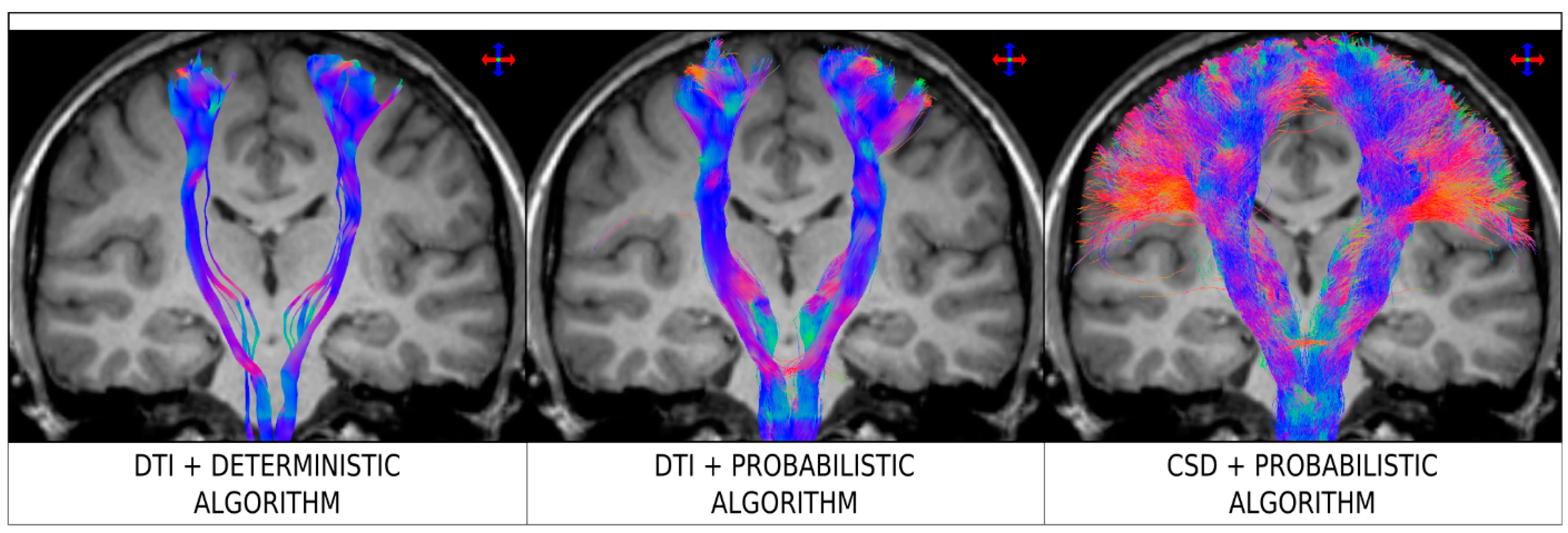


Diagnostics Free Full Text The Seven Deadly Sins Of Measuring Brain Structural Connectivity Using Diffusion Mri Streamlines Fibre Tracking Html
This produced individual subject level maps of brain regions correlating with the seed ROIs Independent mixedeffects group analyses between opioid exposed and controls were conducted for each amygdala seed region using infant sex and maternal depression as covariates as both these factors are shown to impact amygdala connectivity We did not include gestational SeedBased RestingState Functional MRI for Presurgical Localization of the Motor Cortex A TaskBased Functional MRIDetermined Seed Versus an AnatomyDetermined Seed Ji Young Lee , MD, 1, 2 Yangsean Choi , MD, 2 Kook Jin Ahn , MD, PhD, 2 Yoonho Nam , PhD, 2 Jin Hee Jang , MD, 2 Hyun Seok Choi , MD, 2 So Lyung Jung , MD, 2 and Bum Soo Kim , MD 2ABSTRACT Tumor and Edema region present in Magnetic Resonance (MR) brain image can be segmented using Optimization and Clustering merged with seedbased region growing algorithm The proposed algorithm shows effectiveness in tumor detection in T1 w, T2 – w, Fluid Attenuated Inversion Recovery and Multiplanar Reconstruction type MR brain


Arxiv Org Pdf 1812


Intrinsic Functional Connectivity As A Tool For Human Connectomics Theory Properties And Optimization Journal Of Neurophysiology
A systematic review of relations between restingstate functionalMRI and treatment response in major depressive disorder Dichter GS, Gibbs D, Smoski MJ Dichter GS, et al J Affect Disord 15 Feb 1; doi /jjad8 Epub 14 Sep 26 J Affect Disord 15 PMID Free PMC article Review The SPEN singleshot MRI is a recently proposed technique which possesses better robustness to the effects of inhomogeneous field and chemical shift In this paper, we present a highresolution reconstruction method, termed SEED, for SPEN images The SEED combines specific aliasing artifacts reducing approach with weighting CSLesion region Then, histogram thresholding is acquired to automate the seeds selection for region growing process The region is iteratively grown by comparing all unallocated neighboring pixels to the seeds The difference between pixel's intensity value and the region's mean is used as the similarity measure


Identifying Resting State Networks From Fmri Data Using Icas By Gili Karni Towards Data Science


Postimplant Dosimetry Of Permanent Prostate Brachytherapy Comparison Of Mri Only And Ct Mri Fusion Based Workflows International Journal Of Radiation Oncology Biology Physics
By contrast, whenever the cortical seed region (M1 core voxel) was located close to the tumour, the use of nTMS to determine the seed voxel led to more plausible tractography results as compared to fMRI (p < 005), especially for the somatotopic handrelated tractsRead Data and Select Seed Point (s) ¶ We first load a T1 MRI brain scan and select our seed point (s) If you are unfamiliar with the anatomy you can use the preselected seed point specified below, just uncomment the line In1) Left Atrium Seed Region Extraction The seed region inside the left atrium is extracted by first finding the heart region and then searching for seed points that belong to the left atrium To segment the heart region, the Otsu's method is applied to I key to convert it into a binary image The largest connected component is chosen as the initial segmentation of the heart


Resting State Fmri An Overview Sciencedirect Topics


Diffusion Tensor Imaging Dti Fiber Tracking Imagilys
The seed region was derived from the activated brain region (PI patients' DC vs healthy controls' DC) by creating a seeded spherical 5mm region of interest (ROI) around the activated center of mass coordinates Later, FC maps were generated by calculating both positive and negative correlations between the ROIs and other brain voxels The region growing method can work efficiently in medical image segmentation if one can guarantee optimal initial seed point and threshold criterion used to stop growing outside a region In seeded region growing method (SRG), seed selection is crucial, often done by hand in medical image processing , 21Region growing 11,12 is a process that group pixels or previously subdivided regions within the image, into a larger main region based on a predefined selection criterion usually having to do with similarity of intensity values To initialize region growing, seeds are placed within the object desired to be segmented
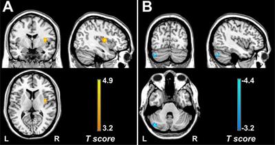


Frontiers Functional Alterations In The Posterior Insula And Cerebellum In Migraine Without Aura A Resting State Mri Study Behavioral Neuroscience
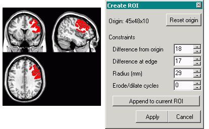


Creating 3d Rois With Mricro
Methods Using 3T functional MRI, we conducted a seedbased restingstate intrinsic FC analysis of the hypothalamus in 18 episodic CH patients during inbout and outofbout periods, and in 19 healthy controls Correlations between hypothalamic FC One of the methods of segmentation the images is the growth method of the area In this study, the region's growth method is used to segment the brain MRI images The method of growing the area consists of several steps In the beginning, you have to select a few initial points (seeds) that are related to the areas to be separated from the fieldThe seedbased functional connectivity analysis was carried out using the Rest software with the left cerebellum (−495,585,185) as the seed region, which was shown to have the greatest difference between ADHD and TDC groups by Zang 10 The time series in the cerebellum were calculated and all voxels were averaged, followed by Pearson correlation analysis with the time


Mriquestions Com Uploads 3 4 5 7 Heuvel Reviewbrainnets1 Pdf


1
3 Seed region growing The basis of the method is to segment an image of N pixels into regions with respect to a set of seeds 26 using only the initial seed pixels The initial seed pixel is selected from a pixel with mask 3X3 These seeds are grown by merging neighboring pixels whose properties are most similar to the To compute functional connectivity maps corresponding to a selected seed region of interest (ROI), the regional time course was correlated against all other voxels within the brain Correlation maps were produced by extracting the BOLD time course from a seed region, then computing the correlation coefficient between that time course and the time course from allSeeded region growing requires seeds as additional input The basic approach is to start with a set of seed points and grow the regions by appending to each seed's neighbouring pixels that have similar properties to the seed The region growing algorithm applied in this study is summarized as follows 1 Histogram calculate histogram of the merged
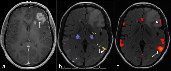


The Role Of Resting State Functional Mri For Clinical Preoperative Language Mapping Cancer Imaging Full Text


Resting State Fmri An Overview Sciencedirect Topics
The segmentation problem was formulated as a problem in region growing In particular, the method started locally by searching for a seed region of the left atrium from an MRI slice A global constraint was imposed by applying a shape prior to the representation of left atrium by Zernike moments 21 Midwest Plains 9U Regional Tournament Seed #5 vs Seed #4 21 Midwest Plains 9U Regional Tournament Seed #5 vs Seed #42 people needed with MRI experience * Full time or 95 – on going role 2 Second postHoliday cover 9th July for 4 weeks To be successful in this role you will possess t


Link Springer Com Content Pdf 10 1007 2f978 3 319 567 2 9077 1 Pdf
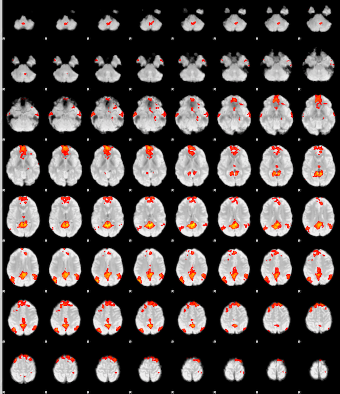


Fsl Fmri Resting State Seed Based Connectivity Neuroimaging Core 0 1 1 Documentation
Seeded Region Growing is an integrated method brought up by Adams and Bischof 413, in which few initial seeds are generated, and more similar neighboring regions are then combined to achieve region growing 1421 In addition, the method of unsupervised vector seeded region growing suitable for medical multispectral images was establishedGroup exponential lasso models were then used to predict gene cluster expression summaries as a function of seed region structural connectivity patterns In several gene clusters, brain regions located in the brain stem, diencephalon, and hippocampal formation were identified that have significant predictive power for these expression summariesSeed selection process Region based segmentation of the medical images is widely used in various clinical applications such as bone and SemiAutomatic Seeded Region Growing for Object Extracted in MRI International Journal of Scientific & Engineering Research Volume 7, Issue 2, February16


3 Dimensional Brain Mri Segmentation Based On Multi Layer Background Subtraction And Seed Region Growing Algorithm Scientific Net


1
Taskbased functional MRI (tbfMRI) is a wellestablished technique used to identify eloquent cortex,but has limitations, particularly in cognitively impaired patients who cannot perform language paradigms Restingstate functional MRI (rsfMRI) is a potential alternative modality for presurgical mapping of language networks thatdoes not require task performance The purpose of our studyFuzzy cmean clustering, seed region growing, and Jaccard similarity coefficient 33 to determine GM and WM tissues in brain MRIs This approach begins by partitioning the given image into several regions The seed region growing method is applied to the image using the centers ofSeedbased functional connectivity analyses and independent components analysis have been used to identify brain regions that show correlated spontaneous lowfrequency BOLD signal fluctuations 37,38 This pattern of intercorrelations over time can then be attributed to discrete, functionally connected brain networks Early skepticism that this technique may be unrelated to significant


Functional Connectivity Mri Studies Experimental Design And Special Applications In Neuroimaging Coursera


Seed Regions Of The Seed Based Resting State Analysis Seed Regions And Download Scientific Diagram


Decreased Hand Motor Resting State Functional Connectivity In Patients With Glioma Analysis Of Factors Including Neurovascular Uncoupling Radiology


Resting State Fmri Wikipedia


Automatic Region Based Brain Classification Of Mri T1 Data
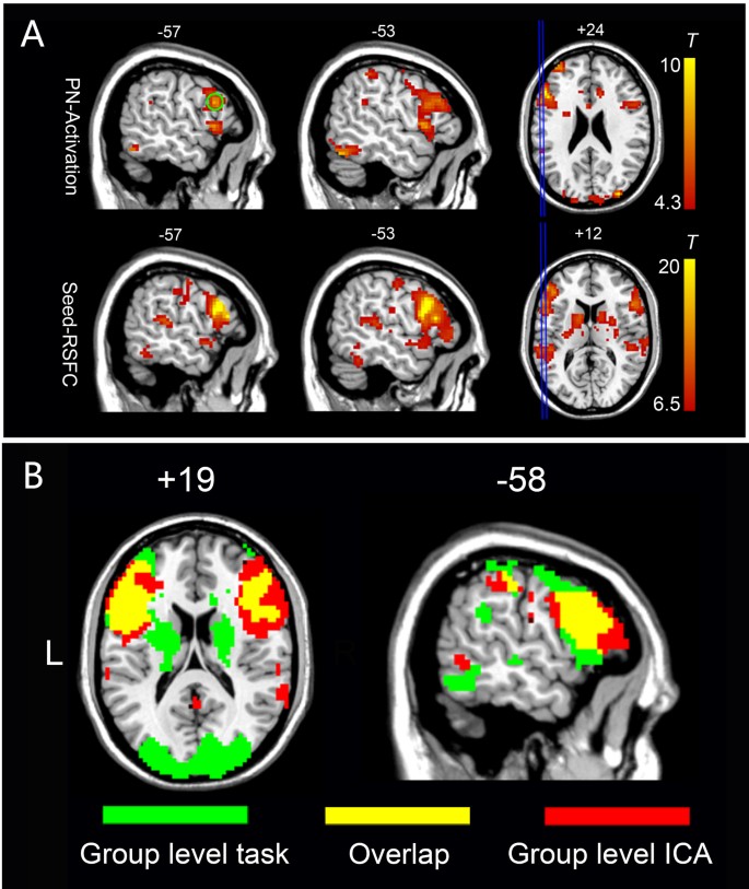


An Automated Method For Identifying An Independent Component Analysis Based Language Related Resting State Network In Brain Tumor Subjects For Surgical Planning Scientific Reports
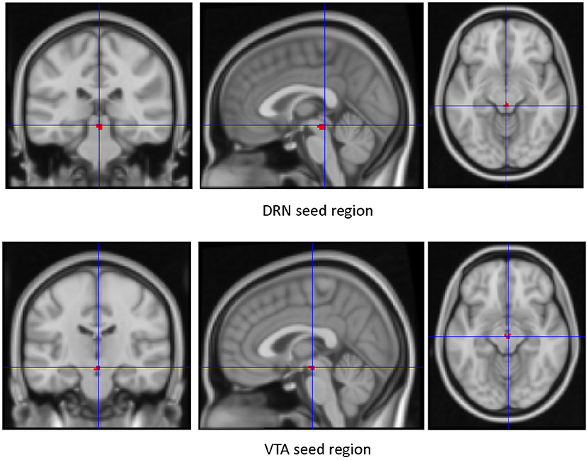


Frontiers Resting State Functional Connectivity Of Dorsal Raphe Nucleus And Ventral Tegmental Area In Medication Free Young Adults With Major Depression Psychiatry


Resting State Functional Mri Everything That Nonexperts Have Always Wanted To Know American Journal Of Neuroradiology
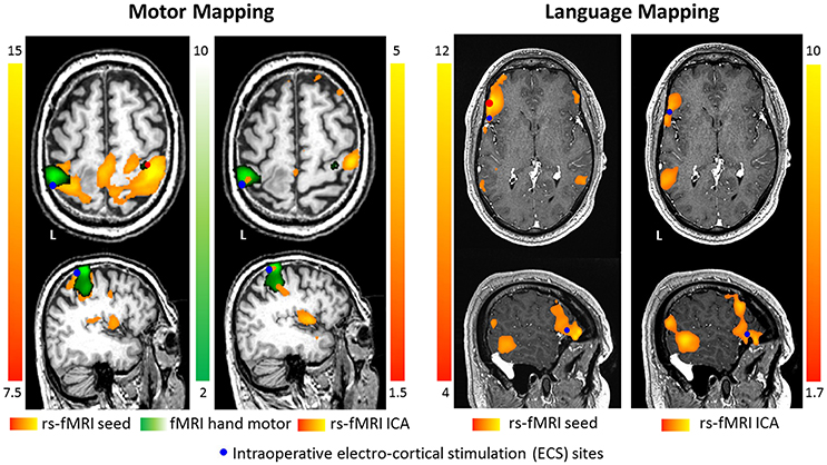


Frontiers Pre Surgical Brain Mapping To Rest Or Not To Rest Neurology
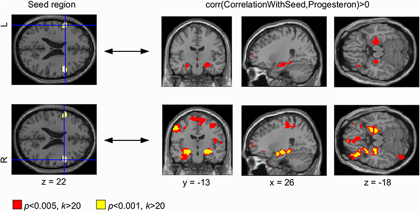


Frontiers Progesterone Mediates Brain Functional Connectivity Changes During The Menstrual Cycle A Pilot Resting State Mri Study Neuroscience


Pineal Region An Approach Radiology Reference Article Radiopaedia Org


Changes In Cerebellar Functional Connectivity And Anatomical Connectivity In Schizophrenia A Combined Resting State Functional Mri And Diffusion Tensor Imaging Study Liu 11 Journal Of Magnetic Resonance Imaging Wiley Online Library
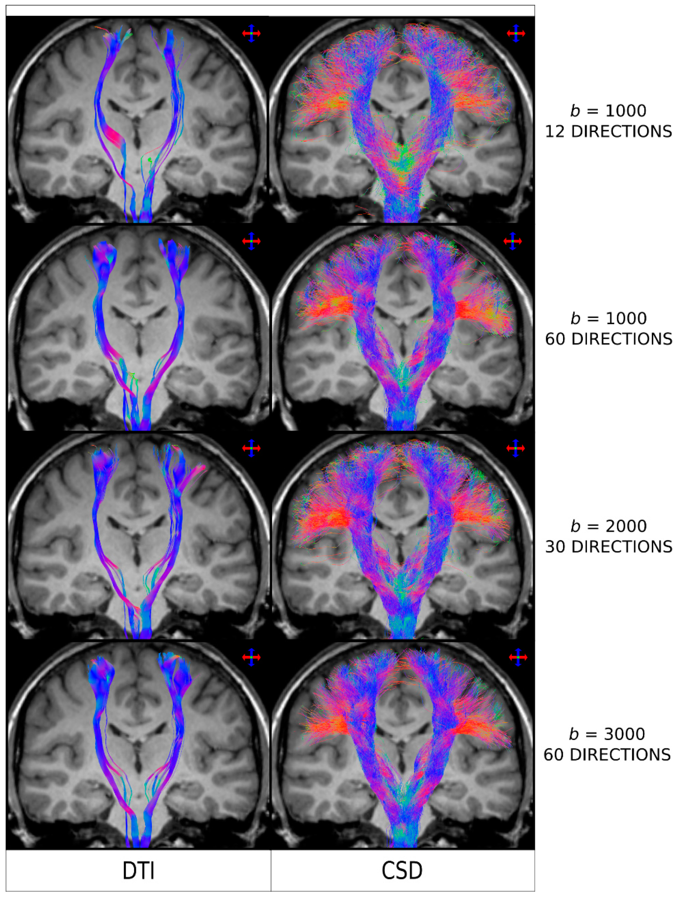


Diagnostics Free Full Text The Seven Deadly Sins Of Measuring Brain Structural Connectivity Using Diffusion Mri Streamlines Fibre Tracking Html


Language Lateralization From Task Based And Resting State Functional Mri In Patients With Epilepsy Rolinski Human Brain Mapping Wiley Online Library


Brain Structure And Function In School Aged Children With Sluggish Cognitive Tempo Symptoms Journal Of The American Academy Of Child Adolescent Psychiatry


Example Of Resting State Modes In The Brain A Activity As Measured Download Scientific Diagram


A Mini Review On Different Methods Of Functional Mri Data Analysis


Fully Balanced Ssfp Without An Endorectal Coil For Postimplant Qa Of Mri Assisted Radiosurgery Mars Of Prostate Cancer A Prospective Study International Journal Of Radiation Oncology Biology Physics


Resting State Seed Based Analysis An Alternative To Task Based Language Fmri And Its Laterality Index American Journal Of Neuroradiology
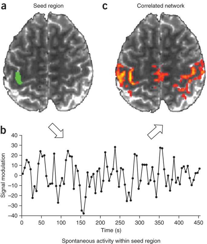


Opportunities And Limitations Of Intrinsic Functional Connectivity Mri Nature Neuroscience


A Novel Magnetic Resonance Imaging Segmentation Technique For Determining Diffuse Intrinsic Pontine Glioma Tumor Volume In Journal Of Neurosurgery Pediatrics Volume 18 Issue 5 16


Figure 3 From A Window Into The Brain Advances In Psychiatric Fmri Semantic Scholar


Anatomical And Functional Organization Of The Human Substantia Nigra And Its Connections Elife


Post Mortem Mapping Of Connectional Anatomy For The Validation Of Diffusion Mri Biorxiv
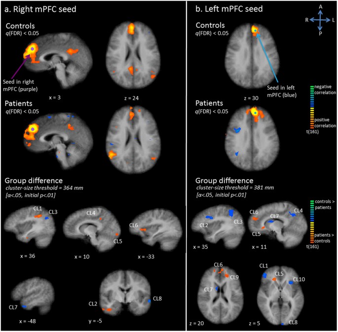


Exploration Of The Brain In Rest Resting State Functional Mri Abnormalities In Patients With Classic Galactosemia Scientific Reports


3


Sleep Deprivation Increases Dorsal Nexus Connectivity To The Dorsolateral Prefrontal Cortex In Humans Pnas


Resting State Functional Mri In Depression Unmasks Increased Connectivity Between Networks Via The Dorsal Nexus Pnas


Ultra High Field Mri Reveals Mood Related Circuit Disturbances In Depression A Systematic Comparison Between 3 Tesla And 7 Tesla Biorxiv


Resting State Functional Mri Everything That Nonexperts Have Always Wanted To Know American Journal Of Neuroradiology


Depiction Of The Bilateral Subthalamic Nucleus Seed Region As Derived Download Scientific Diagram


Exploration Of The Brain In Rest Resting State Functional Mri Abnormalities In Patients With Classic Galactosemia Scientific Reports


The Human Brain Is Intrinsically Organized Into Dynamic Anticorrelated Functional Networks Pnas


Altered Hypothalamic Functional Connectivity In Cluster Headache A Longitudinal Resting State Functional Mri Study Journal Of Neurology Neurosurgery Psychiatry
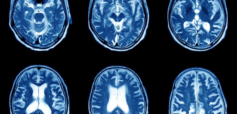


Early Mris May Predict 9 Year Outcomes In Ms Children Study Says


Resting State Fmri Wikipedia


Maps Of Seed Based Resting State Fmri Functional Connectivities The Download Scientific Diagram


Changes In Thalamus Connectivity In Mild Cognitive Impairment Evidence From Resting State Fmri European Journal Of Radiology


Performing Region Growing On An Mri Bone Image A Original Images Download Scientific Diagram
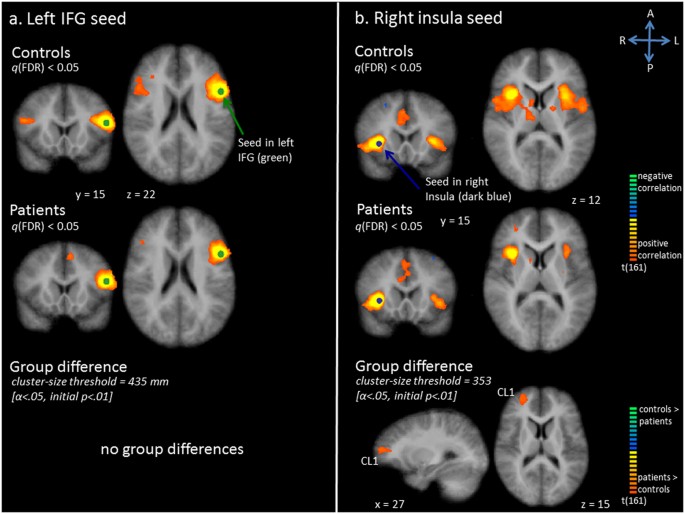


Exploration Of The Brain In Rest Resting State Functional Mri Abnormalities In Patients With Classic Galactosemia Scientific Reports
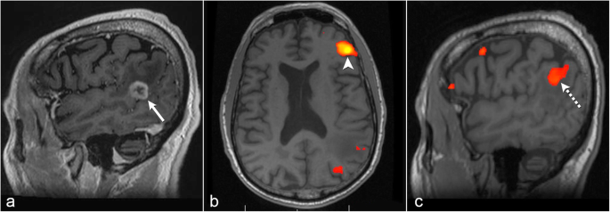


The Role Of Resting State Functional Mri For Clinical Preoperative Language Mapping Cancer Imaging Full Text


The Role Of Resting State Functional Mri For Clinical Preoperative Language Mapping Cancer Imaging Full Text


Advanced Brain Tumour Segmentation From Mri Images Intechopen
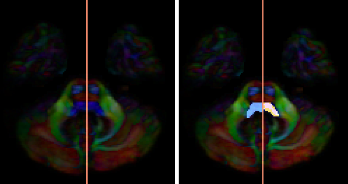


Medial Lemniscus Ml Whole Brain Protocol For Tractography With Empirical Mri
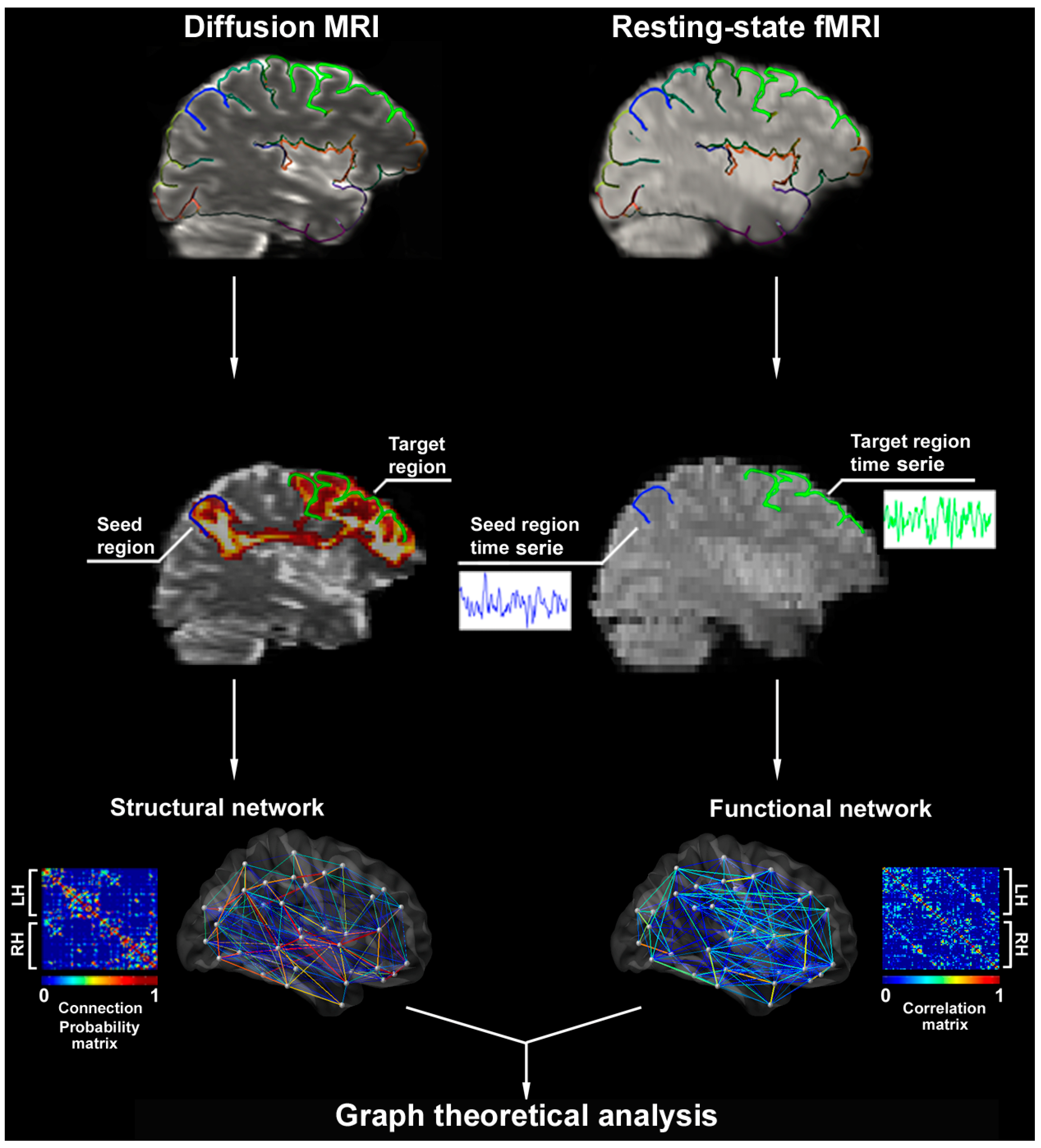


Brain Sciences Free Full Text Contributions Of Imaging To Neuromodulatory Treatment Of Drug Refractory Epilepsy Html


Magnetism Questions And Answers In Mri


Specific Connectivity With Operculum 3 Op3 Brain Region In Acoustic Trauma Tinnitus A Seed Based Resting State Fmri Study Biorxiv
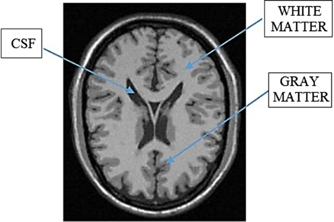


Automatic Seeded Region Growing Asrg Using Genetic Algorithm For Brain Mri Segmentation Springerlink
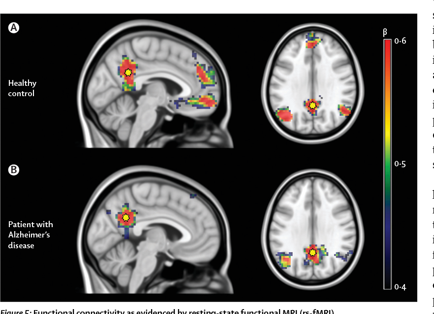


Figure 5 From Multimodal Imaging In Alzheimer S Disease Validity And Usefulness For Early Detection Semantic Scholar


Resting State Functional Mri Everything That Nonexperts Have Always Wanted To Know American Journal Of Neuroradiology


The Potential Of Multimodal Mri For Understanding Sports


Functional Mri Vs Navigated Tms To Optimize M1 Seed Volume Delineation For Dti Tractography A Prospective Study In Patients With Brain Tumours Adjacent To The Corticospinal Tract Sciencedirect


Restoring Susceptibility Induced Mri Signal Loss In Rat Brain At 9 4 T A Step Towards Whole Brain Functional Connectivity Imaging


Presurgical Resting State Functional Mri Language Mapping With Seed Selection Guided By Regional Homogeneity Hsu Magnetic Resonance In Medicine Wiley Online Library
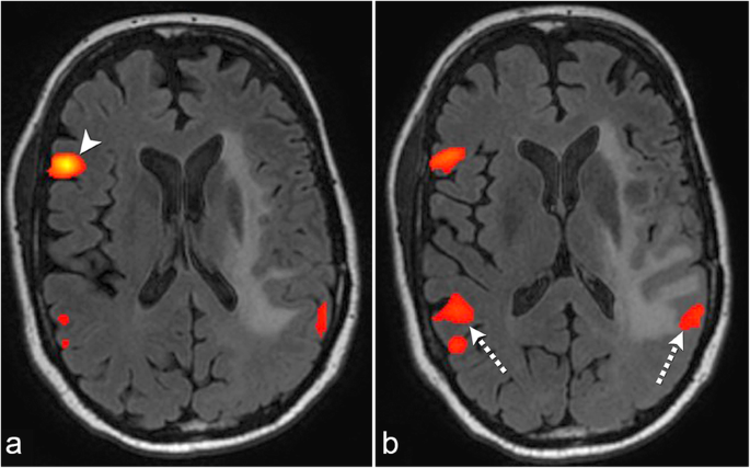


The Role Of Resting State Functional Mri For Clinical Preoperative Language Mapping Cancer Imaging Full Text


The Role Of Resting State Functional Mri For Clinical Preoperative Language Mapping Cancer Imaging Full Text


Physicists Engineers To Build Next Generation Mri Brain Scanner


Resting State Seed Based Analysis An Alternative To Task Based Language Fmri And Its Laterality Index American Journal Of Neuroradiology


Characterizing The Spectrum Of Task Fmri Connectivity Approaches Ppt Download


Resting State Functional Mri Everything That Nonexperts Have Always Wanted To Know American Journal Of Neuroradiology


Resting State Functional Magnetic Resonance Imaging Connectivity Between Semantic And Phonological Regions Of Interest May Inform Language Targets In Aphasia Journal Of Speech Language And Hearing Research


High Connectivity Between Reduced Cortical Thickness And Disrupted White Matter Tracts In Long Standing Type 1 Diabetes Diabetes


Altered Regional And Circuit Resting State Activity In Patients With Occult Spastic Diplegic Cerebral Palsy Pediatrics Neonatology


How To Safely Perform Magnetic Resonance Imaging Guided Radioactive Seed Localizations In The Breast Journal Of Clinical Imaging Science


Real Time Presurgical Resting State Fmri In Patients With Brain Tumors Quality Control And Comparison With Task Fmri And Intraoperative Mapping Abstract Europe Pmc


2
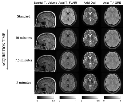


Ismrm Digital Posters Page Interventional Interventional Mr And Safety Issues


Main Brain Areas Implicated In Resting State Functional Mri Diagram Of Download Scientific Diagram


Looking At Connections Between Brain Regions 1 Some


Figure 2 From Structural And Functional Mri Study Of The Brain Cognition And Mood In Long Term Adequately Treated Hashimoto S Thyroiditis Semantic Scholar
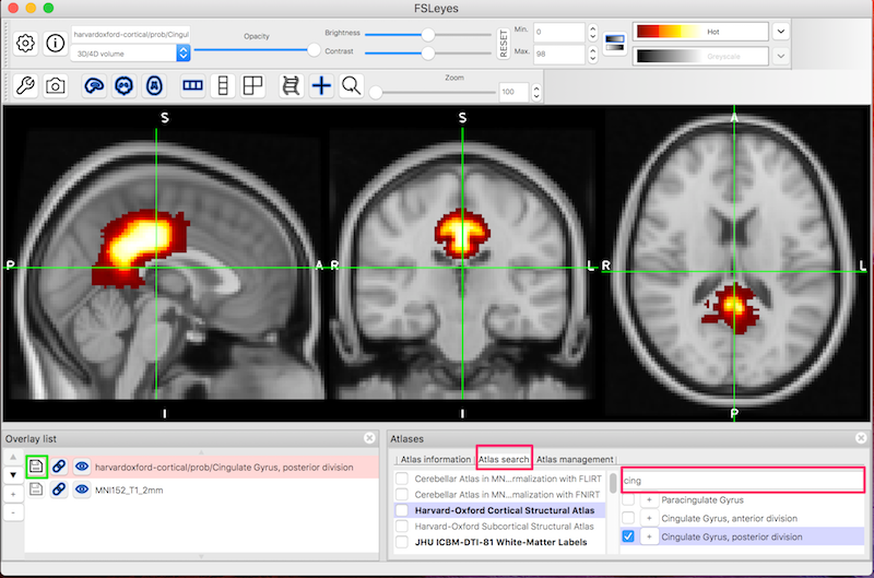


Fsl Fmri Resting State Seed Based Connectivity Neuroimaging Core 0 1 1 Documentation


A Functional Magnetic Resonance Imaging Study On The Neural Mechanisms Of Hyperalgesic Nocebo Effect Journal Of Neuroscience


Diffusion Tensor Imaging Dti Fiber Tracking Imagilys


Concepts And Principles Of Clinical Functional Magnetic Resonance Imaging Chapter 13 The Cambridge Handbook Of Research Methods In Clinical Psychology
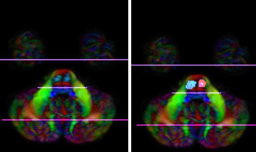


Corticospinal Tract Cst Whole Brain Protocol For Tractography With Empirical Mri


Altered Regional And Circuit Resting State Activity In Patients With Occult Spastic Diplegic Cerebral Palsy Pediatrics Neonatology


Q Tbn And9gctasix9izbbdou Vuspvet3lzbqgtuf9uveuz4wv5i3qds4ev4 Usqp Cau
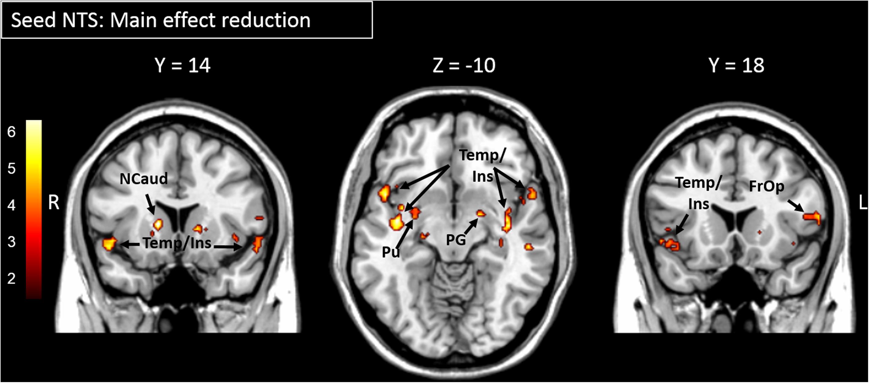


Frontiers Functional Connectivity Within The Gustatory Network Is Altered By Fat Content And Oral Fat Sensitivity A Pilot Study Neuroscience


Resting State Functional Mri Everything That Nonexperts Have Always Wanted To Know American Journal Of Neuroradiology
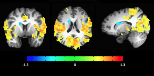


Mri Resting State Fmri Carney Institute For Brain Science Brown University


Cef Seed Regions Displayed On Coronal And Sagittal Views Of The Download Scientific Diagram


Correlation Of Copper Measurements With Functional Connectivity Mri Download Scientific Diagram


0 件のコメント:
コメントを投稿